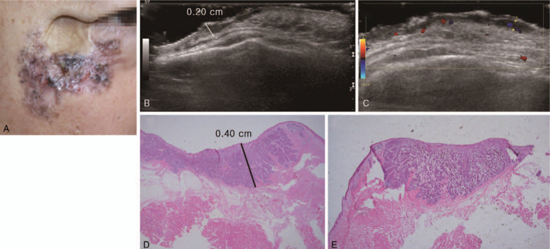
Figure 1.

A 65-year-old male patient with basal cell carcinoma on the right cheek and lower eyelid. The patient received a split thickness skin graft on the right cheek 30 years ago due to trauma. (A) Clinical photography. Irregular margined skin lesion with multiple pigmentation and crater-like appearance on the right cheek and lower eyelid. (B) Ultrasonography longitudinal scan. (C) Ultrasonography transverse scan. 4.0 × 3.0 × 0.2 cm sized hypoechoic lesion along the skin and subcutaneous fat layer. (D and E) Histopathologic image. The thickness of basal cell carcinoma was 0.40 cm (hematoxylin and eosin stain, magnification 1.25×).
