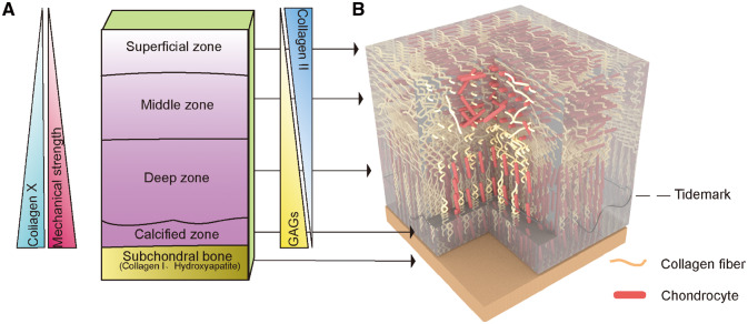
Figure 1.
The five different layers of AC show zone-specific cell morphologies, matrix compositions, collagen fibril orientations and mechanical properties. (A) The content of collagen type X and GAG and compressive strain increase with depth, while the collagen type II concentration is inversely proportional to depth in the cartilaginous region. Subchondral bone is composed mainly of collagen type I and hydroxyapatite. (B) (1) The superficial zone has the highest density of chondrocytes and collagen fibres parallel to the joint surface. (2) The Middle zone has randomly oriented collagen fibres. (3) The deep zone has fibres perpendicular to the joint surface and tidemark, which is a basophilic line between uncalcified and calcified cartilage. (4) The calcified zone is the transition from cartilage to bone, and it has hypertrophic chondrocytes and anchors the fibres to the subchondral bone

