Figure 8.

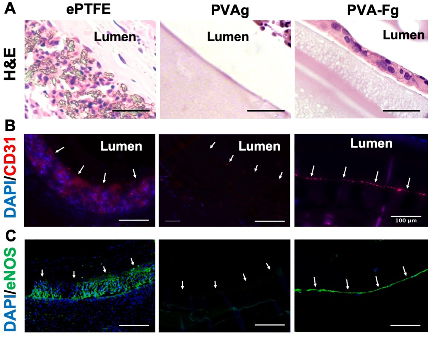
In-vivo endothelialization of PVA-Fg grafts. (A) Hematoxylin and eosin (H&E) staining of middle section of explanted vascular grafts. PVA-Fg showed a layer of cells on the luminal surfaces. Scale bar = 50 μm. (B) Immunofluorescence staining of the middle section of explanted vascular grafts. Arrows indicate luminal surfaces. Scale bar = 100 μm. CD31 positive signals in PVA-Fg samples indicated the presence of endothelial cells. (C) Immunofluorescence staining of the middle section of explanted vascular grafts. Arrows indicate luminal surfaces. Scale bar = 100 μm. The eNOS positive signals indicated that the endothelial cells inside PVA-Fg samples expressed eNOS proteins.
