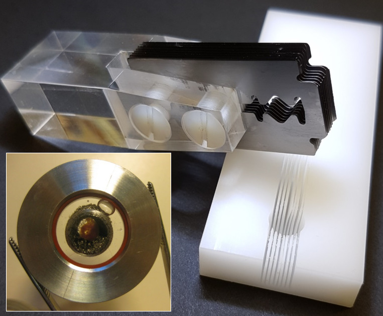
Figure 1.
Preparation of ex vivo tissue slices. The matrix cutting block (right) has an 8-mm-diameter hemispherical cavity with eight slots, 0.2 mm wide (on 1-mm centers). An isolated heart is placed into the cavity, and a set of eight razor blades (0.1-mm thick, spaced 1 mm apart, screwed to a Plexiglas handle; top left), is inserted into matching slots in the cutting block so the heart can be cut into multiple sections in one stroke. The assembled imaging chamber (inset) contains a cardiac tissue slice on the lower coverslip, held in place by a nylon mesh stretched across a stainless steel tambour.

