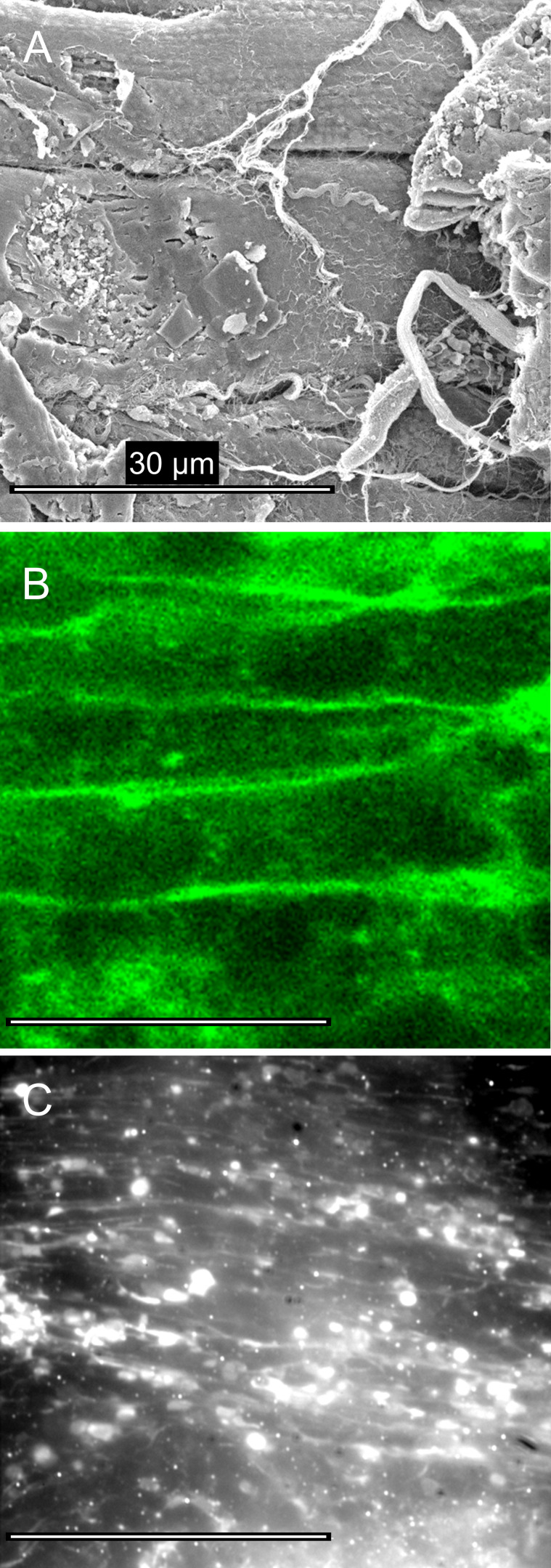
Figure 2.
Tissue morphology after the preparation of ex vivo heart slices. (A) Scanning electron micrograph of a critical-point dried cardiac tissue section from adult mouse shows regions of damage where myocytes are cut obliquely (right side) and other regions where muscle cell plasma membrane is relatively intact (see text for details). (B) Scanning confocal optical microscopy, using the membrane dye (DeepRed Cellmask, 10 µg ⋅ ml−1, for 10 min; Invitrogen) shows that the cellular structures in optical sections taken 1 µm beyond the cut surface were well-preserved. (C) Tissue sections, labeled with Cy3-telenzepine, viewed by TIRFM, were heterogeneous. Some regions were well-preserved while others showed significant cell damage. Bright circular regions occur where the plasma membrane is closely opposed to the coverslip surface and enters the evanescent field.

