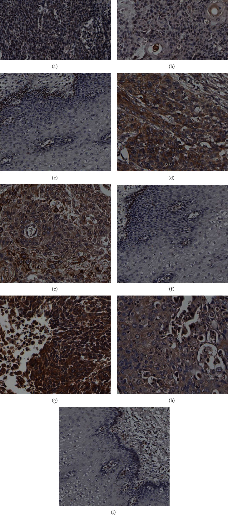
Figure 1.

Immunohistochemical staining of FGF2, FGFR3, FGFBP1 expression in ESCC and normal esophagus mucosa. (a, b) Positive expression of FGF2 in ESCC tumor tissue; (c) positive expression of FGF2 in the basal layer of normal esophageal mucosa; (d, e) positive expression of FGFR3 in ESCC tumor tissue; (f) positive expression of FGFR3 in the basal layer of normal esophageal mucosa; (g, h) positive expression of FGFBP1 in ESCC tumor tissue; (i) negative expression of FGFBP1 in normal esophageal mucosa.
