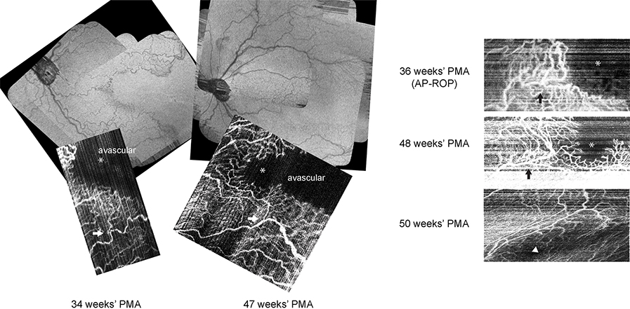
FIG 1.
Optical coherence tomography (OCT) retina views and longitudinal OCT angiography imaging of the perifoveal microvasculature in a preterm infant at 34, 36, 47, 48, and 50 weeks’ postmenstrual age (PMA) who developed aggressive posterior retinopathy of prematurity at 36 weeks’ PMA and received intravitreal bevacizumab injection. At 34 weeks’ PMA, the vascular pattern was consistent with temporal notch without closure of temporal perifoveal vasculature. At 36 weeks’ PMA, vascular growth stalled, with dilated shunt vessels across the nasal macula and at the vascular margin. At 47 and 48 weeks’ PMA, perifoveal vessels and capillaries both were extending beyond the prior vascular-avascular junction, with less dilation and tortuosity than previously seen; the decreased rim of perifoveal shunting allowed growth of capillary branches toward the location of the impending fovea. At 50 weeks’ PMA, complete formation of the perifoveal vasculature (with motion artifact) was seen. Black and white arrows mark the corresponding retinal microvasculature location. Asterisks marks the possible location of impending fovea; the arrowhead marks the fovea.

