
Figure 2.
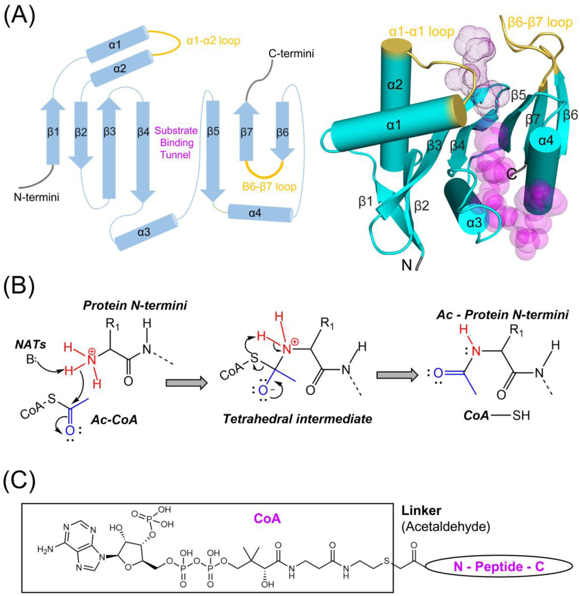
Some NATs function independently, either in monomeric or homodimer form. (A) Structures of monomeric StRimI (PDB: 2CNM), SsNAT (PDB: 4LX9), uncomplexed SpNAA10 (PDB: 4KVX), hNAA50 (PDB: 3TFY), hNAA40 (PDB: 4U9W), and DmNAA80 (PDB: 5WJE) are shown in cartoon and color in cyan. The α1- α2 and β6-β7 substrate binding loops are highlighted in yellow. Substrate in the structures are shown in stick and colored in magenta. The hNAA40-specific N terminal domain is highlighted in grey. (B) Dimeric RimL (PDB:1S7N) and hNAA60 (PDB: 5ICW) are shown. The α1- α2 and β6-β7 substrate binding loops are highlighted in yellow. Substrate in the structures are shown in stick and colored in magenta. To form a dimer, StRimL utilizes the two β6 strands from each subunit, while hNAA60 uses the extended β6- β7 loops. The NAA60-specific N terminal domain is highlighted in grey. NAT catalytic subunits that have not been shown to function independently are not shown.