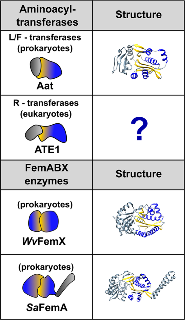
Figure 3.
GNAT folds of the FemABX enzymes and the aminoacyl tRNA transferases. The left-hand column depicts a cartoon representation of each protein, with domains that contain the GNAT fold colored with a blue-yellow gradient. The right-hand column contains ribbon depictions of the three-dimensional structures (if known). The α-helices and β-strands comprising the GNAT fold are colored blue and yellow, respectively. For clarity, only one of the GNAT folds is colored in the FemABX structures. PDB IDs are as follows: L/F transferase (2DPS); Weissella viridescens FemX (1P4N); and Staphylococcus aureus FemA (1LRZ). Notable is the lack of any three-dimensional structure of an ATE1 (R-transferase).

