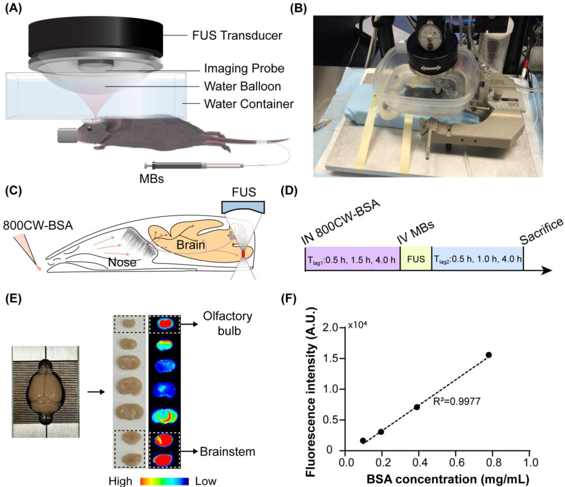
Figure 1.

Experimental methods. (A) A schematic and (B) picture of the FUS system for mouse brain treatment. Illustration of the (C) FUSIN procedure and (D) experimental timeline. IN delivery of 800CW-BSA was followed by FUS treatment targeting at the brainstem. (E) After FUSIN treatment, the mouse brain was harvested and cut into 2-mm coronal slices for fluorescence imaging. The high fluorescence intensity observed at the olfactory bulb confirmed that IN-administered 800CW-BSA transport to the brain along the olfactory pathway. (F) 800CW-BSA fluorescence intensity at different dilutions showed a linear relationship between fluorescence intensity and the concentration of 800CW-BSA.
