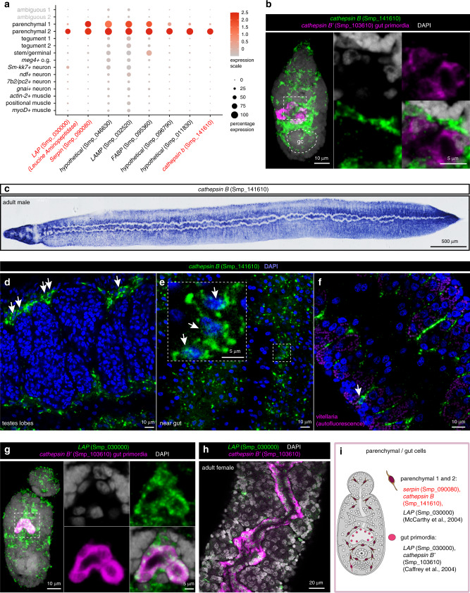
Fig. 4. Identification of schistosome parenchymal and primordial gut cells.
a Expression profiles of cell marker genes that are specific or enriched in the parenchymal clusters. Genes validated by ISH are marked in red. b Double FISH of parenchymal cathepsin B (Smp_141610) with a known marker of differentiated gut, cathepsin B’ (Smp_103610), MIP. No expression of parenchymal cathepsin B is observed in the primordial gut. gc: germinal cell cluster. c WISH of parenchymal cathepsin B in adult males. d–f FISH of parenchymal cathepsin B in different regions of adult worms: d testes lobes, e gut, and f vitellaria. White arrows indicate positive cells. Single confocal sections shown. g, h lap (Smp_030000) is expressed in both parenchyma and in the g gut primordia as well as h adult gut, shown by double FISH with the gut cathepsin B (Smp_103610). i Schematic that summarises the parenchymal cell populations in 2-day schistosomula. Marker genes identified in the current study are indicated in red. All previously reported genes are shown in black. The numbers of ISH experiments performed for each gene are listed in ‘Methods’ and Supplementary Data 7.

