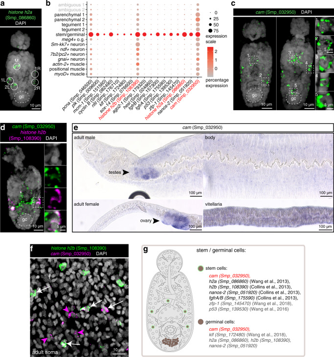
Fig. 5. A single cluster of stem cells in 2-day old schistosomula.
a FISH of h2a (Smp_086860) shows ~5 stem cells located at distinct locations—1 medial cell (M) and 2 lateral cells on each side (1L and 2L, 1R, and 2R; L: left; R: right), MIP. b Expression profiles of cell marker genes that are specific or enriched in the stem/germinal cell cluster. Genes validated by ISH are marked in red. c FISH of cam (Smp_032950) shows a similar localisation pattern as h2a, with some worms with a few more cam+ cells in the medial region as well as in the germinal cell cluster region, MIP. gc: germinal cluster. d Double FISH of cam (Smp_032950) and a previously validated schistosome stem cell marker h2b (Smp_108390), MIP. e WISH of cam (Smp_032950) in adult parasites shows enriched expression in the gonads including testis, ovary, and vitellarium, as well as in the mid-animal body region. f Double FISH of cam and h2b in adult soma. A single confocal section is shown. White arrows indicate co-localisation of two genes and magenta arrowheads indicate cells expressing only cam. g Schematic that summarises the stem and germinal cell populations in 2-day old schistosomula. Marker genes identified in the current study are indicated in red. All previously reported genes are shown in black. Genes that are enriched in this cluster but have not been directly shown by ISH are shown in grey. The numbers of ISH experiments performed for each gene are listed in ‘Methods’ and Supplementary Data 7.

