Figure 1.
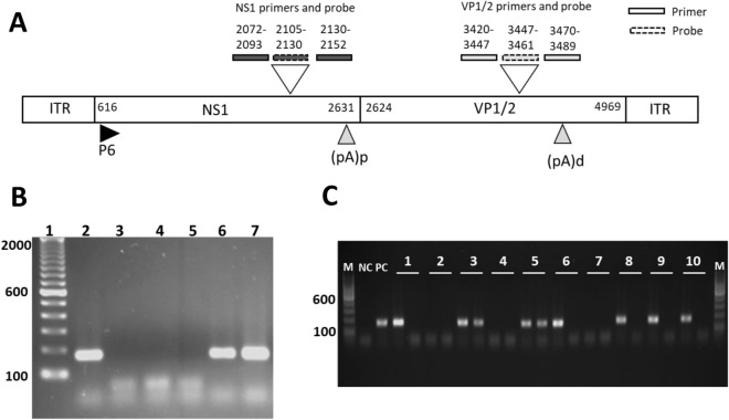
(A) Schematic description of the B19V genome organization and PCR design. Gene locus and primer and probe localization for NS1 and VP1/2 are indicated by numbers representing the nucleotide position. ITR inverted terminal repeat, NS1 non structural protein 1, VP1/2 capsid proteins, P6 P6 promotor, (pA)p polyadenylation site proximal, (pA)d polyadenylation site distal. (B) Representative agarose gel electrophoresis gel blot image of a B19V-VP1/2 DNA and RNA-positive EMB using VP1/2 specific nested-PCR. Amplicon length 173 bps. 1 = DNA-Marker 100 bps; 2 = positive control; 3 = negative control; 4 = PCR after DNA extraction and DNAse treatment; 5 = PCR after RNA extraction, RNAse treatment and RT-PCR; 6 = PCR after DNA extraction; 7 = PCR after RNA extraction and DNAse treatment and RT-PCR. Complete gel blot image of figure (B) was shown in Supplementary Fig. S1. (C) Representative agarose gel electrophoresis gel blot image of 10 EMB samples following VP1/2 specific nested PCR. DNA (first lane) and cDNA (second lane) of each EMB were analysed. Amplicon length 173 bps EMBs 1, 6, 8, 9 and 10 were tested positive for viral DNA and negative for viral RNA. EMBs 3 and 5 were positive for both, viral RNA and DNA. EMBs 2, 4 and 7 were virus negative without any viral DNA nor RNA being detectable. M 100 bps marker, NC negative control, PC positive control.

