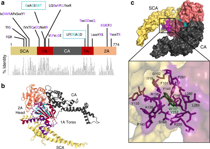
Fig. 3. Structure and sequence conservation of HelD.
a Linear representation of the structure of HelD with a map of coloured domains and conserved sequence motifs (top), and a histogram of sequence identity (bottom). b The structure of HelD (bottom) coloured according to the schematic in panel a. SCA is shown in yellow, SF1 helicase-like domain 1 A in red, CA in dark grey, and SF1 helicase-like domain 2 A in orange. Conserved sequence motifs are shown in purple, with the Walker A/B ATP-binding site in cyan. c, shows a surface-rendered impression of HelD with the same colouring as in b. The bottom panel shows an expanded view of the boxed region in the top panel with a semi-transparent surface and conserved amino acids that form the ‘Trp cage’ shown as purple sticks, and the conserved Trp in dark green.

