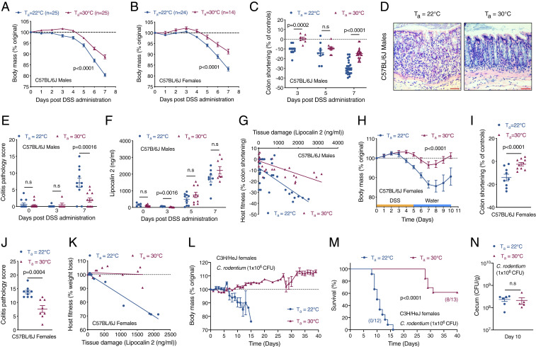
Fig. 1.
Thermoneutral mice exhibit intestinal disease tolerance. (A–G) To induce colitis, C57BL/6J mice housed at Ta = 22 °C or Ta = 30 °C were administered 4% DSS in drinking water for 7 d. (A–C) Body mass of male (A) and female (B) C57BL/6J treated mice (n = 25 per temperature for males and n = 14 to 25 per temperature for females; data pooled from n = 4 independent experiments and analyzed by two-way ANOVA) and colon shortening in C57BL/6J male treated mice (n = 7 to 25 per time point and temperature; data pooled from n = 4 independent experiments and analyzed by Student’s t test). (D) Representative pictures of hematoxylin and eosin (H&E)-stained sections of colon. (Scale bar: 100 μM.) (E) Colitis pathology score in C57BL/6J male treated mice (n = 6 to 15 per time point and temperature; data pooled from n = 3 independent experiments and analyzed by Student’s t test). (F) Plasma levels of lipocalin 2, a tissue damage marker, in C57BL/6J male mice administered DSS and housed at Ta = 22 °C or Ta = 30 °C (n = 5 to 11 per time point and temperature; data pooled from n = 3 independent experiments and analyzed by Student’s t test). (G) Reaction norm plots of host fitness (colon shortening) and tissue damage (lipocalin 2) during DSS-induced colitis in C57BL/6J male mice housed at Ta = 22 °C or Ta = 30 °C (n = 35 per temperature; data pooled from n = 4 independent experiments). (H–K) To monitor tissue regeneration, mice were administered 4% DSS in drinking water for 5 d followed by recovery over the next 5 d. Body mass (H; analyzed by two-way ANOVA), colon shortening (I), and colitis pathology score (J) of C57BL/6J female treated mice (n = 9 to 10 per temperature; data pooled from n = 2 independent experiments and analyzed by Student’s t test). (K) Reaction norm plots of host fitness (weight loss) and tissue damage (lipocalin 2) during DSS-induced colitis and recovery in female C57BL/6J mice housed at Ta = 22 °C or Ta = 30 °C (n = 9 to 10 per temperature; data pooled from n = 2 independent experiments). (L–N) C3H/HeJ female mice housed at Ta = 22 °C or Ta = 30 °C were orally infected with C. rodentium [1 × 10⁶ colony-forming units (CFUs)]. Body mass (L) and survival (M) was monitored over 40 d (n = 12 to 13 per temperature; data pooled from n = 2 independent experiments; survival curves analyzed by log-rank Mantel–Cox test). (N) Bacterial CFUs in cecum were quantified on day 10 (n = 5 to 6 per temperature; data representative of n = 2 independent experiments and analyzed by Student’s t test). Data are presented as mean ± SEM.

