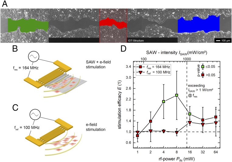
Fig. 3.
SAW stimulation of the ectodermal cell line MDCK-II. (A) High-quality micrograph image of the progressive in vitro wound-healing in a confluent monolayer at t = 5 h. The colored regions indicate the SAW-stimulated cells (red) and the internal references (green and blue). (B and C) Representations of the different stimuli mechanism at different frequencies. While there is a simultaneous mechanical and electrical stimulation at the SAW resonance frequency, we depict in B the result of an electrical stimulus only. This is achieved by detuning the IDT frequency to a somewhat lower value outside its bandwidth, where no SAW is excited (C). (D) Power dependency on SAW stimulation at different frequencies. There is significant improvement of cell growth and migration rate up to 135 ± 85% for SAW-supported cell growth at PIN = 4 and 8 mW. Exceeding ISAW > 1 W/cm2 leads to a decrease of stimulation efficacy E. A significant increase of the efficacy (P < 0.05) compared to an external low control is indicated by the color of the symbols’ inner area. The median time for surface coverage with MDCK-II cells is about 31 h.

