Fig. 1.
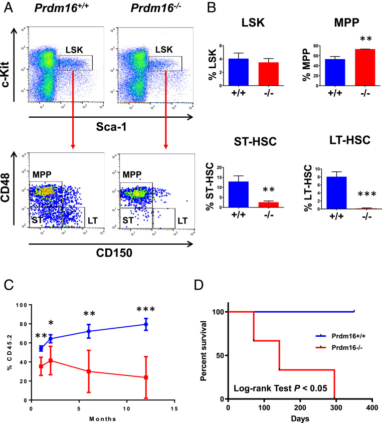
Significant reduction in fetal liver HSCs after Prdm16 deletion. (A) Representative FACS analysis of LSK compartments including LT-HSC (LT), ST-HSC (ST), and MPP in fetal livers of e18.5 Prdm16−/− versus Prdm16+/+ embryos. (B) Quantification of results in A (n = 3 for each group). The frequencies (mean ± SD) of LSK cells in total bone marrow nucleated cells and of LT-HSC/ST-HSC/MPP populations in LSK cells are shown. (C) Competition of 1 × 10⁶ e18.5 Prdm16−/− or Prdm16+/+ fetal liver cells against equal number of wild-type C57BL/6-Ly5.2 bone marrow cells in lethally irradiated C57BL/6-Ly5.2 recipient mice. The frequencies (mean ± SD) of fetal liver cells (CD45.2+) in peripheral blood of recipient mice at 1, 2, 6, and 12 mo after transplantation are shown (n = 5 for each fetal liver genotype). (D) Survival curves of irradiated C57BL/6-Ly5.2 mice receiving 1 × 10⁶ bone marrow cells from primary recipient mice of e18.5 Prdm16−/− or Prdm16+/+ fetal liver cells (n = 3 for each genotype). *P < 0.05; **P < 0.01; ***P < 0.001.

