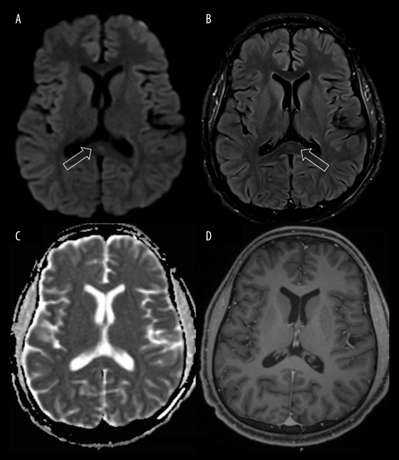
Figure 1.
Brain Magnetic resonance imaging (MRI) on admission. Diffusion-weighted (A) and fluid-attenuated inversion recovery (B) imaging demonstrates a hyperintense signal in the splenium of corpus callosum, with associated loss of signal on apparent diffusion coefficient maps (C) corresponding to restricted diffusion. T1-weighted images with contrast (D) showed an isointense signal without contrast enhancement. These findings were suggestive of cytotoxic lesion of corpus callosum. Arrows indicate the splenium of the corpus callosum.

