Figure 4.

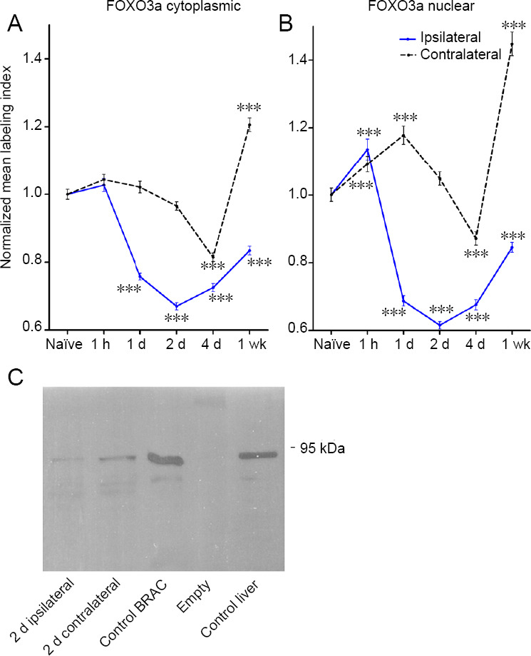
Alterations in forkhead class box O3a (FOXO3a) expression in response to nerve injury and antibody specificity control.
Cytoplasmic (A) and nuclear (B) FOXO3a immunofluorescence relative mean intensity levels ± SEM observed in dorsal root ganglion (DRG) neurons ipsilateral and contralateral to injury at time points as indicated. Each graph point represents a quantitative analysis of n = 441–663 individual neurons analyzed/experimental time point and condition with three animals/ experimental time point and condition analyzed (18 animals in total). ***P < 0.001 (Kruskal-Wallis one-way analysis of variance with Dunn's post hoc test analysis signifies significant difference from the naïve state). (C) Western blot analysis of anti-FOXO3a treated membrane of electrophoresed protein extracts from 2 days-injured ipsilateral L4–6 DRG (2 days ipsilateral), 2 days-injured contralateral L4–6 DRG (2 days contralateral), control BrCA1 (BRCA) cell line, and control rat liver. Western blots were performed in triplicate. Note: The FOXO3a antibody recognizes a band of approximately 85–90 kDa, consistent with the expected molecular weight for FOXO3a and nerve injury results in reduced levels of FOXO3a. Empty refers to the lane at which no sample was loaded.
