Figure 5.

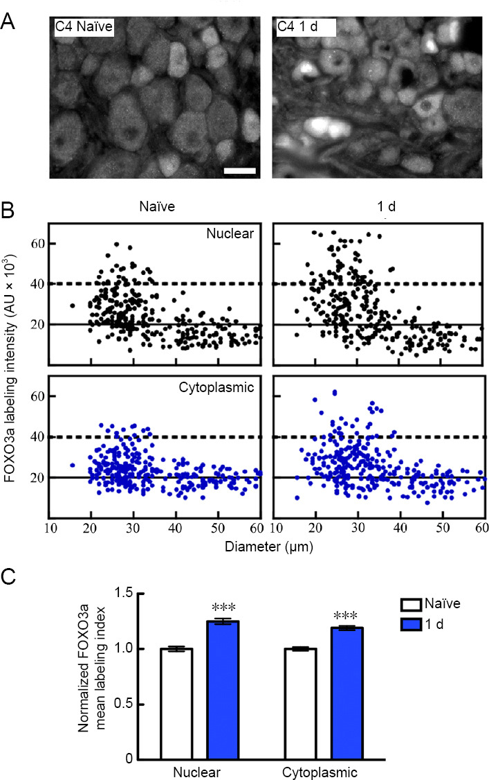
L4–6 spinal nerve transection alters DRG neuronal FOXO3a expression and localization in uninjured C4 ganglia remote from the injury site.
(A) FOXO3a immunofluorescence in C4 DRG sections collected from naïve rats and the rats that underwent 1 day unilateral L4–6 spinal nerve injury. Scale bar: 50 μm. Note: L4–6 spinal nerve injury results in elevated nuclear and cytoplasmic FOXO3a staining in small- to medium-sized neurons of the uninjured C4 DRG. (B) Scatterplots that plot point data representing the relationship between the FOXO3a nuclear (top) and cytoplasmic (bottom) labelling index and cell body diameter. Solid lines divide the plots into the least labelled and moderately labelled populations; dotted lines separate the moderately labelled from heavily labelled populations of FOXO3a expressing neuros. (C) Bar graphs representing normalized nuclear (left) and cytoplasmic (right) FOXO3a mean labeling index ± SEM. (***P < 0.0001, vs. naïve; Mann-Whitney U test). Each graph bar represents a quantitative analysis of n = 667–708 individual neurons analyzed/experimental condition from three animals in total per condition. DRG: Dorsal root ganglion; FOXO3a: forkhead class box O3a.
