Figure 1.
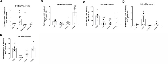
Dysregulated DRs mRNA levels in BV-2 microglial cells exposed to PD mimetics (LPS, protenone and 6-OHDA) after 24 hours.
BV-2 cells at low passage (< 25) were plated at a density of 1.5 × 10⁵ cells in 6-well plates with 10% FGM until 80% confluent and then incubated for further 24 hours in the presence of either LPS (1 μg/μL), rotenone (0.01 μM) and 6-OHDA (25 μM) in 1% FGM. mRNA was quantified using the ΔCt method and normalized using the 18S gene. Data are expressed as the mean ± SEM, obtained from two independent determinations, each run in duplicate or triplicate (n = 4–6). Bar graphs depicting mRNA levels of (A) the dopamine D1 receptor, (B) the dopamine D2 receptor, (C) the dopamine D3 receptor, (D) the dopamine D4 receptor and (E) the dopamine D5 receptor. *P < 0.05, **P < 0.01 vs. control (one-way ANOVA followed by a Sidak post hoc test). 18S: Ribosomal protein 18S (housekeeping gene); 6-OHDA: 6-hydroxydopamine; DR: dopamine receptor; LPS: lipopolysaccharide; PD: Parkinson's disease; Ref: reference.

