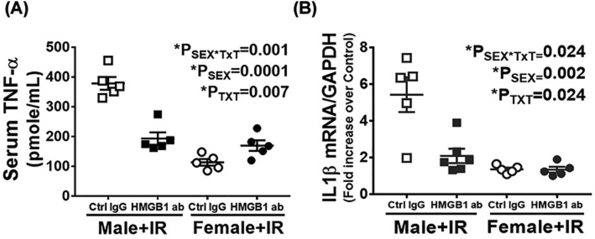
Figure 3. Neutralizing HMGB1 attenuates IR-induced increases in inflammatory cytokine levels 24 h post-IR only in male SHR.

Serum and kidney samples were collected 24 ht-IR in 13 week old male and female SHR treated with control IgG or HMGB1 neutralizing antibody 1 h or to IR. Serum TNF-α was quantified by ELISA (A) and renal IL1β mRNA expression was measured by RT-PCR (B). Data are expressed as means ± SEM; n=5–6 rats in each group with individual animal data indicated by the symbols. Open symbols indicate control IgG-treated animals, filled symbols indicate anti-HMGB1-treated animals, males are represented by squares and females by circles. Data were compared via two-way ANOVA with P<0.05 considered significant.
