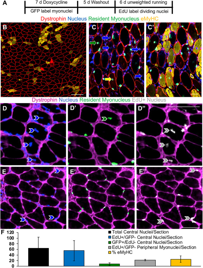
Figure 4.

Soleus myonuclear dynamics during unweighted wheel running. (A) Study design illustrating resident myonuclear labelling in HSA‐GFP mice, ethenyldeoxyuridine (EdU) labelling for DNA synthesis, and unweighted wheel running. (B) Representative image of immunohistochemistry (IHC) for dystrophin (red) and embryonic myosin heavy chain (eMyHC, orange), scale bar = 100 μm. (C) Representative image of IHC for dystrophin (red), nuclei (DAPI), and green fluorescent protein (GFP)‐labelled non‐satellite cell‐derived resident myonuclei (green), with eMyHC (orange) added in (C’). In (C), blue arrows point to central nuclei found in eMyHC+ fibres, the green arrow points to a GFP+ central myonucleus in an eMyHC− fibre, and the yellow arrow points to a GFP+ myonucleus in an eMyHC+ fibre. (D–D”) Representative IHC images showing a GFP+/EdU− myonucleus (green arrow) and GFP−/EdU+ central myonuclei (grey arrows) within dystrophin (pink). (E–E”) Representative images showing EdU+ peripheral myonuclei (grey arrows). (F) Prevalence of total central nuclei, EdU+/GFP− central nuclei, GFP+/EdU− central nuclei, EdU+/GFP− peripheral myonuclei, and eMyHC expression. Scale bars = 50 μm, n = 4 for running and n = 3 for sedentary (data not shown for sedentary mice due to negligble EdU and eMyHC expression).
