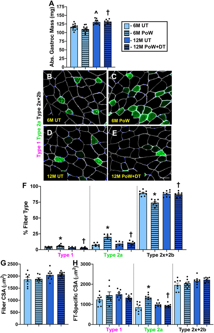
Figure 5.

Gastrocnemius muscle size and fibre type with training and detraining. (A) Absolute gastrocnemius muscle wet weight in 6M UT, 6M PoW, 12M UT, and 12M PoW + DT. (B–E) Repesentative immunohistochemistry images from the four groups showing dystrophin (white), type 2a (green), unstained type 2x and/or 2b fibres (black), and DAPI (blue). (F) Fibre type (FT) distribution. (G) Average muscle fibre cross‐sectional area (CSA). (H) Fibre type‐specific CSA. *6M UT versus 6M PoW, ^6M UT versus 12M UT, #12M DT versus 12M PoW + DT, †6M PoW versus 12M PoW + DT. All P < 0.05, scale bars = 50 μm, n ≥ 8 per group for each analysis.
