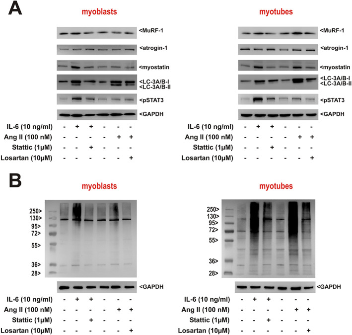
Figure 8.

Role of interleukin‐6 (IL‐6) and angiotensin II (Ang II) pathway activation in rat skeletal myoblasts and myotubes in vitro. (A) Rat skeletal muscle cells (RSkMC) myoblasts (left panel) or RSkMC myotubes (right panel) were treated with Stattic (1 μM) or losartan (10 μM) for 1 h before adding IL‐6 (10 ng/mL) or Ang II (100 nM). Homogenates of RSkMC were probed with antibodies detecting muscle‐specific RING finger 1 (MuRF1), atrogin‐1, myostatin, light chain 3 (LC3) (isoform A + B), and pSTAT3. GAPDH served as a loading control. (B) RSkMC myoblasts (left panel) or RSkMC myotubes (right panel) were treated as described previously and probed with an antibody detecting ubiquitin. A molecular weight marker to estimate the size of the ubiquitinated proteins is included. GAPDH served as a loading control. (A, B) n = 8 samples per group, four independent experiments.
