Figure 4.

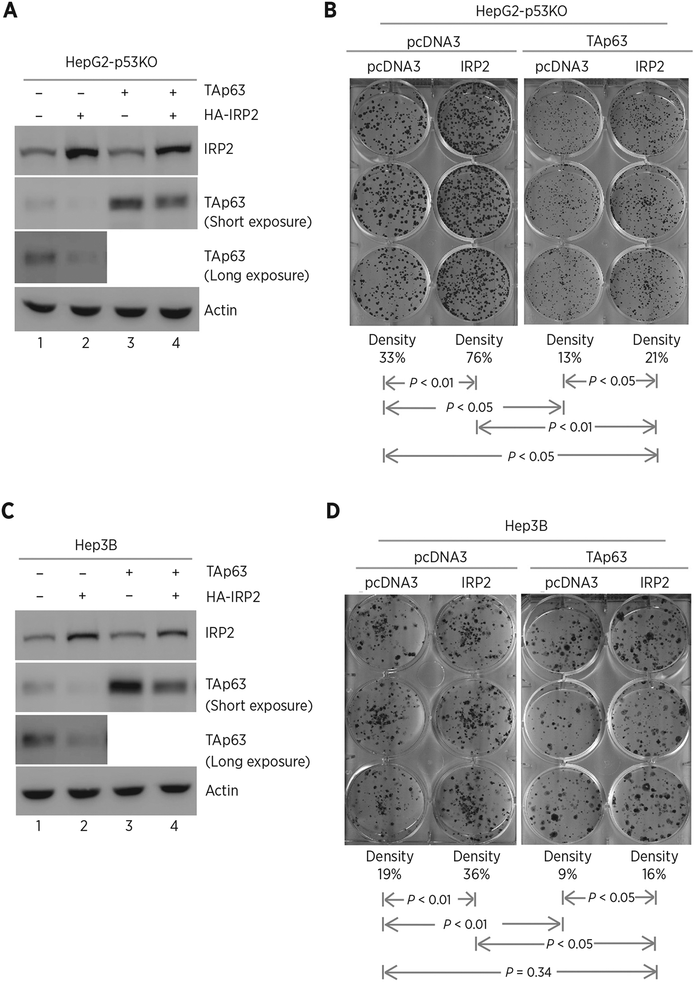
Ectopic expression of IRP2 promotes cell growth via TAp63. A, p53−/− HepG2 cells were transfected with control pcDNA3 or a vector expressing HA-tagged IRP2 along with a control vector or a vector expressing TAp63. Twenty-four hours posttransfection, cell lysates were collected and subjected to Western blot analysis to detect IRP2, TAp63, and actin proteins. B, Colony formation assay was performed with p53−/− HepG2 cells transfected with control pcDNA3 or a vector expressing HA-tagged IRP2, followed by cotransfection with a control vector or a vector expressing TAp63. The relative density for colonies was showed below each image. C and D, The experiments were performed as in A and B except that p53−/− Hep3B cells were used.
