Figure 6.

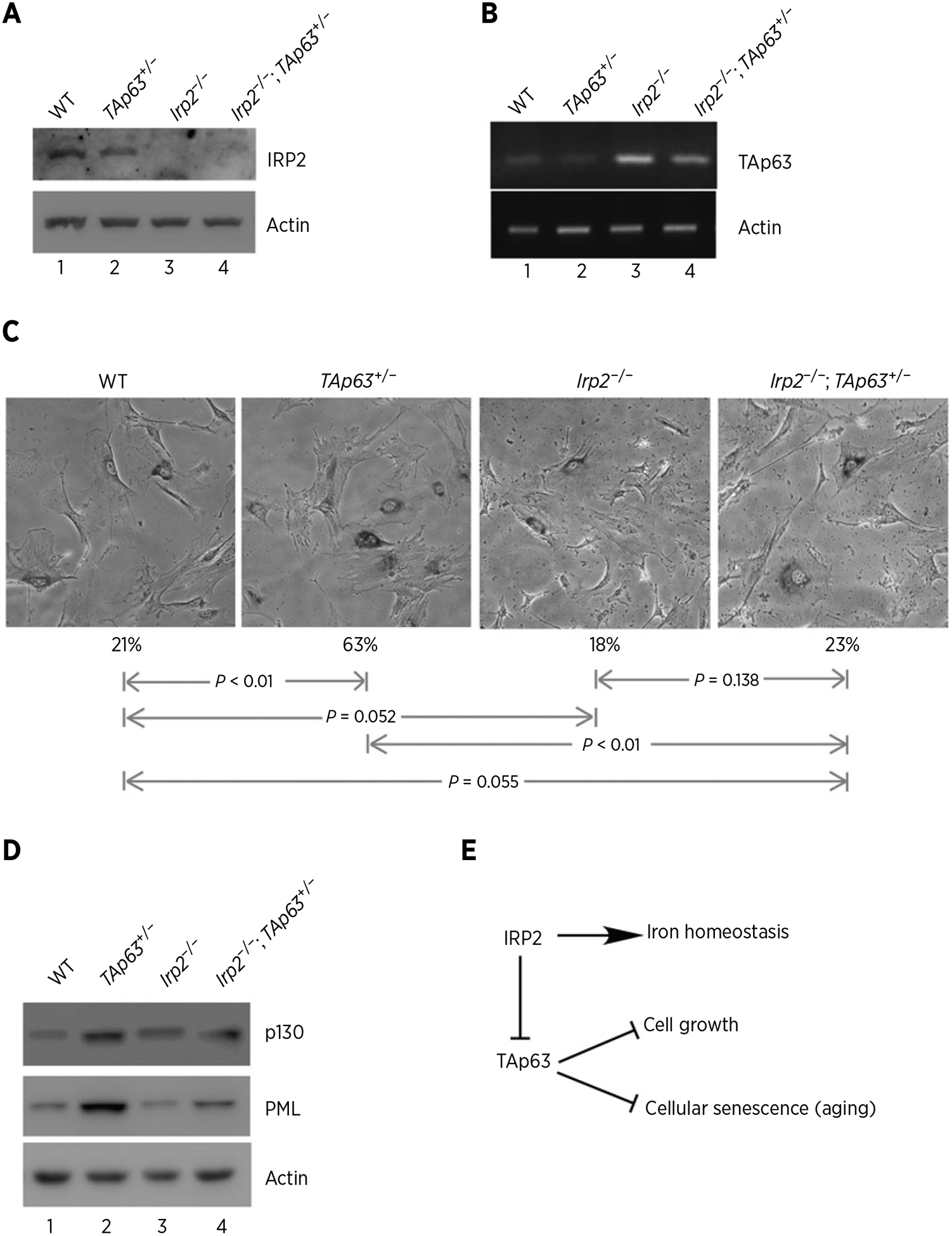
Loss of IRP2 inhibits cellular senescence induced by TAp63 deficiency. A, Western blots were prepared using extracts from WT, TAp63+/−, Irp2−/−, and Irp2−/−;TAp63+/− littermate MEFs. The blots were probed with antibodies against Irp2 and actin. B, The levels of p63 and actin transcripts were measured in WT, TAp63+/−, Irp2−/−, and Irp2−/−;TAp63+/− MEFs. C, Representative images of SA-β-Gal–stained WT, TAp63+/−, Irp2−/−, and Irp2−/−;TAp63+/− MEFs. Quantification of the percentage of SA-β-Gal–positive cells was shown below each image. D, Western blots were prepared using extracts from WT, TAp63+/−, Irp2−/−, and Irp2−/−;TAp63+/− MEFs. The blots were probed with antibodies against p130, PML and actin, respectively. E, A model of how IRP2 modulates cell growth and cellular senescence via TAp63.
