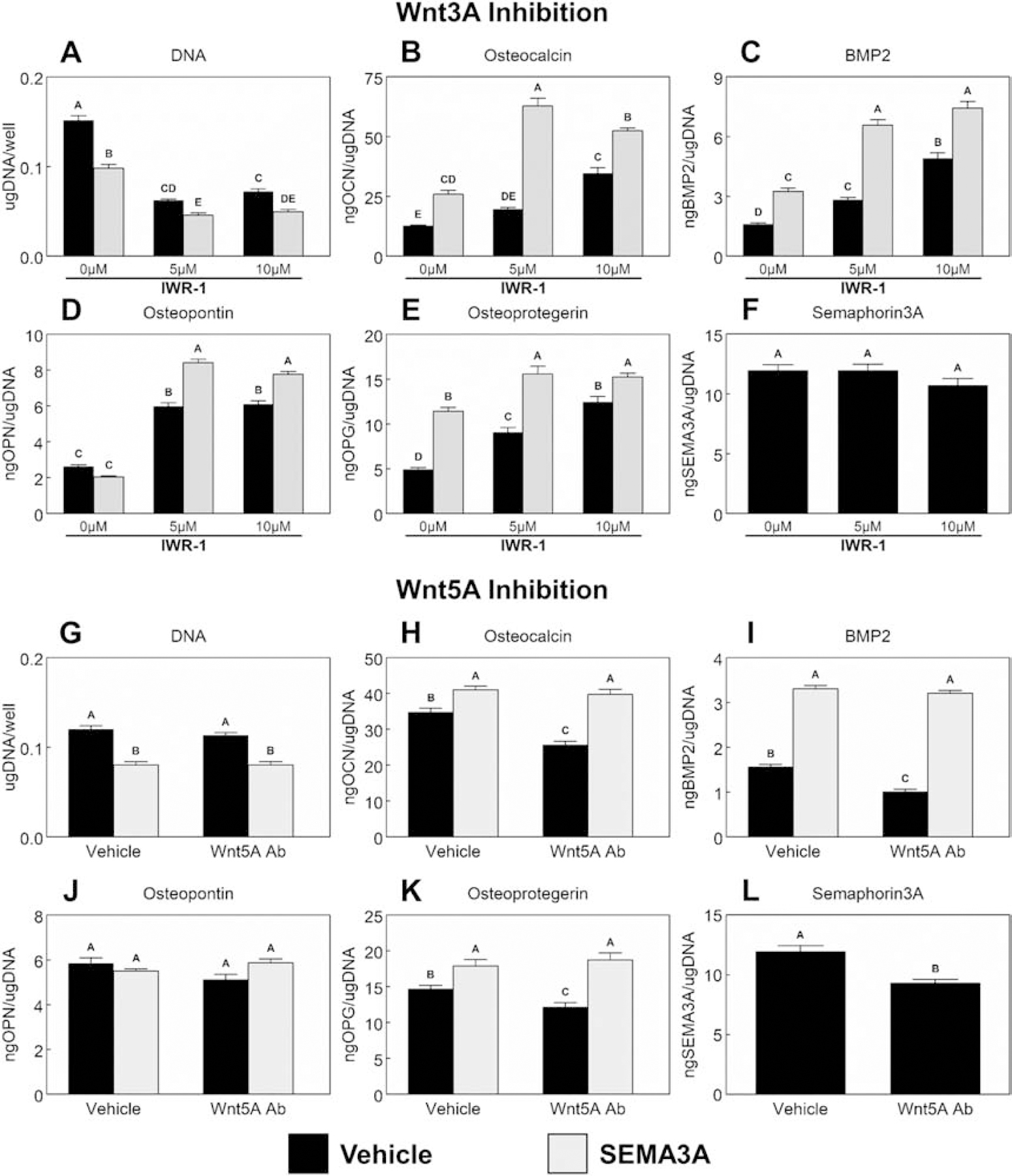
Figure 4. Effect of Wnt signaling and Sema3A on MSC response to microstructured and hydrophilic surfaces.

MSCs were cultured on TCPS or Ti substrates. Cultures supplemented with or without 1μg/mL Sema3A were treated with either 5μM or 10μM IWR-1 (A – F) to inhibit Wnt3A signaling or a 1:200 dilution of a polyclonal anti-Wnt5A antibody (G – L) to inhibit Wnt5A signaling for 7d. Cells were then treated with fresh media for 24h. After 24h, media were collected, and cell lysates were assayed for DNA content (A, G). Media were assayed for osteocalcin (B, H), BMP2 (C, I), osteopontin (D, J), osteoprotegerin (E, K) and Semaphorin3A (F, L). Data shown are the mean ± standard error (SE) of six independent samples. Groups not sharing a letter are statistically significant at α=0.05.
