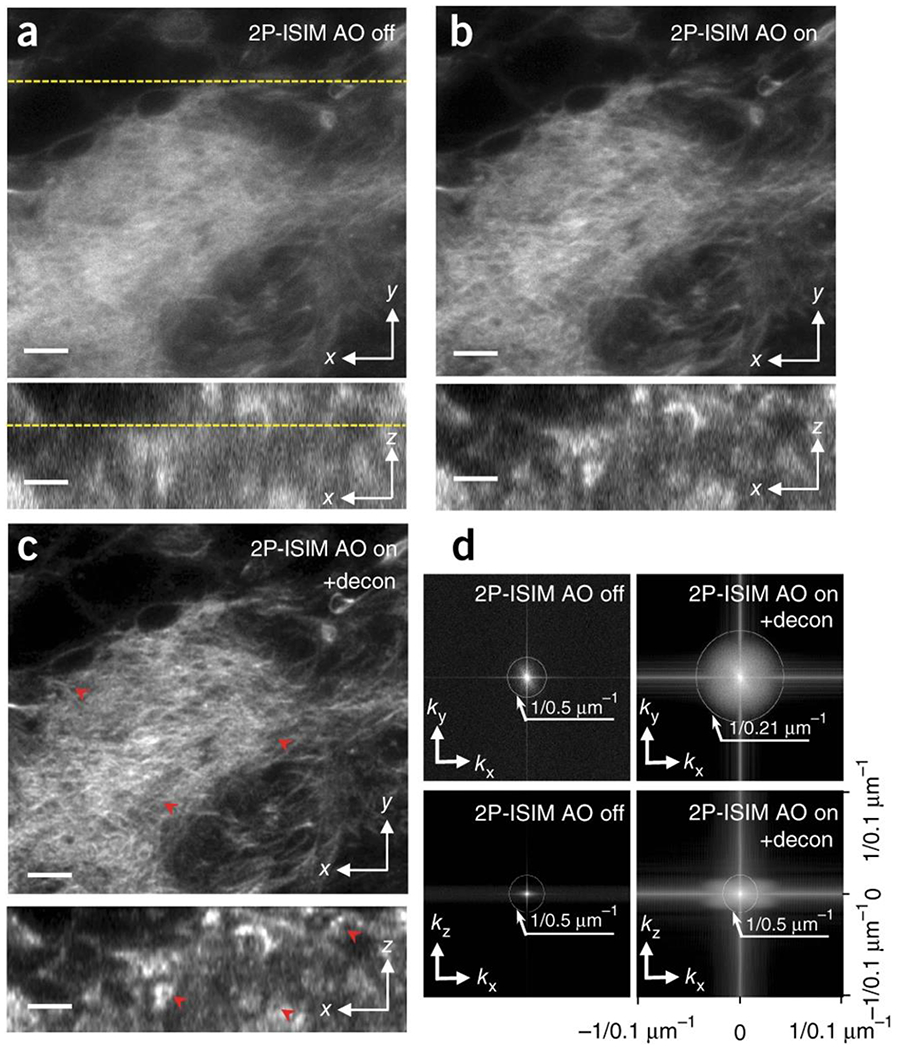
Fig. 1. AO correction based on direct wavefront sensing improves spatial resolution of 2P-ISIM in biological samples, as revealed in labeled Drosophila third instar larval brain.

Lateral (top) and axial (bottom) 2P ISIM images of Alexa Fluor 488 phalloidin labeled actin in fixed larval brain lobe, shown without (a) and with (b) adaptive correction, and after subsequent deconvolution (c). The lateral slice is taken 35 μm from the surface of the brain lobe; correspondence with axial slice is indicated with yellow dotted line. Progressive improvements in spatial resolution and contrast are evident in a-c (see also red arrowheads), and further quantified in d), where lateral (top row) and axial (bottom row) MTFs are shown. Note that axial resolution is limited to the step size used when acquiring stacks (0.5 μm for this dataset). See also Supplementary Fig. 6, Supplementary Video 1. Scale bars: 5 μm.
