Figure 1.

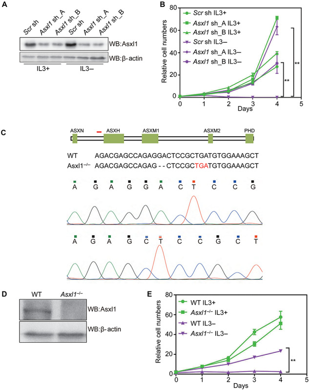
Depletion of Asxl1 confers IL3-independent growth of 32D cells. (A) WB analysis of Asxl1 in control (Scr sh) and Asxl1-KD 32D cells (Asxl1 sh_A and Asxl1 sh_B) in the presence or absence of IL3. (B) Cell growth curves of 32D cells expressing the indicated shRNAs under culture conditions with or without IL3. MTS assay was employed at the specified time points to follow the rate of proliferation. (C) The schematic showing nucleotide deletion of Asxl1 by the CRISPR/Cas9 method. The short red line marks the location of designed sgRNA. The premature stop codon is highlighted in red in the below sequences. Sequencing peaks around the sgRNA docking site is shown at the bottom. (D) WB analysis of total protein lysates of WT and Asxl1-KO 32D cells. β-actin served as a loading control. (E) Cell growth curves of WT and Asxl1-KO 32D cells under culture conditions with or without IL3. Significance level was determined using Student’s two-sided t-tests. The error bars denote SD, n = 3; **P ≤ 0.01.
