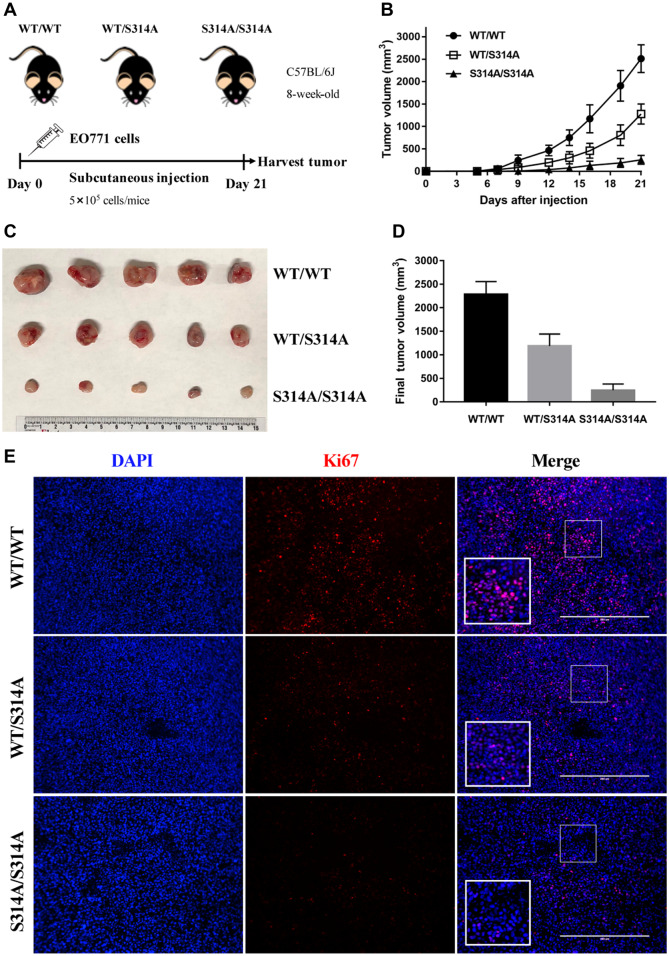
Figure 1.
MdmxS314A mice exhibit enhanced tumor control. (A) Experimental design scheme. EO771 cells were injected into the right flank of mice (n = 5/genotype) on Day 0 and tumors were harvested on Day 21. Tumor size was measured by caliper every other day. (B) Growth curve of EO771 syngeneic tumors in different genotypic mice over 21 days. (C) Gross appearance of tumors derived from EO771 cells on Day 21 after implantation into wild-type, MdmxWT/S314A, and MdmxS314A/S314A mice. (D) Mean values of tumor volume in different genotypic mice on Day 21. (E) Immunofluorescence for Ki67 (red) and DAPI (blue) in EO771 tumor sections from wild-type, MdmxWT/S314A, and MdmxS314A/S314A mice. Scale bar, 400 μm.

