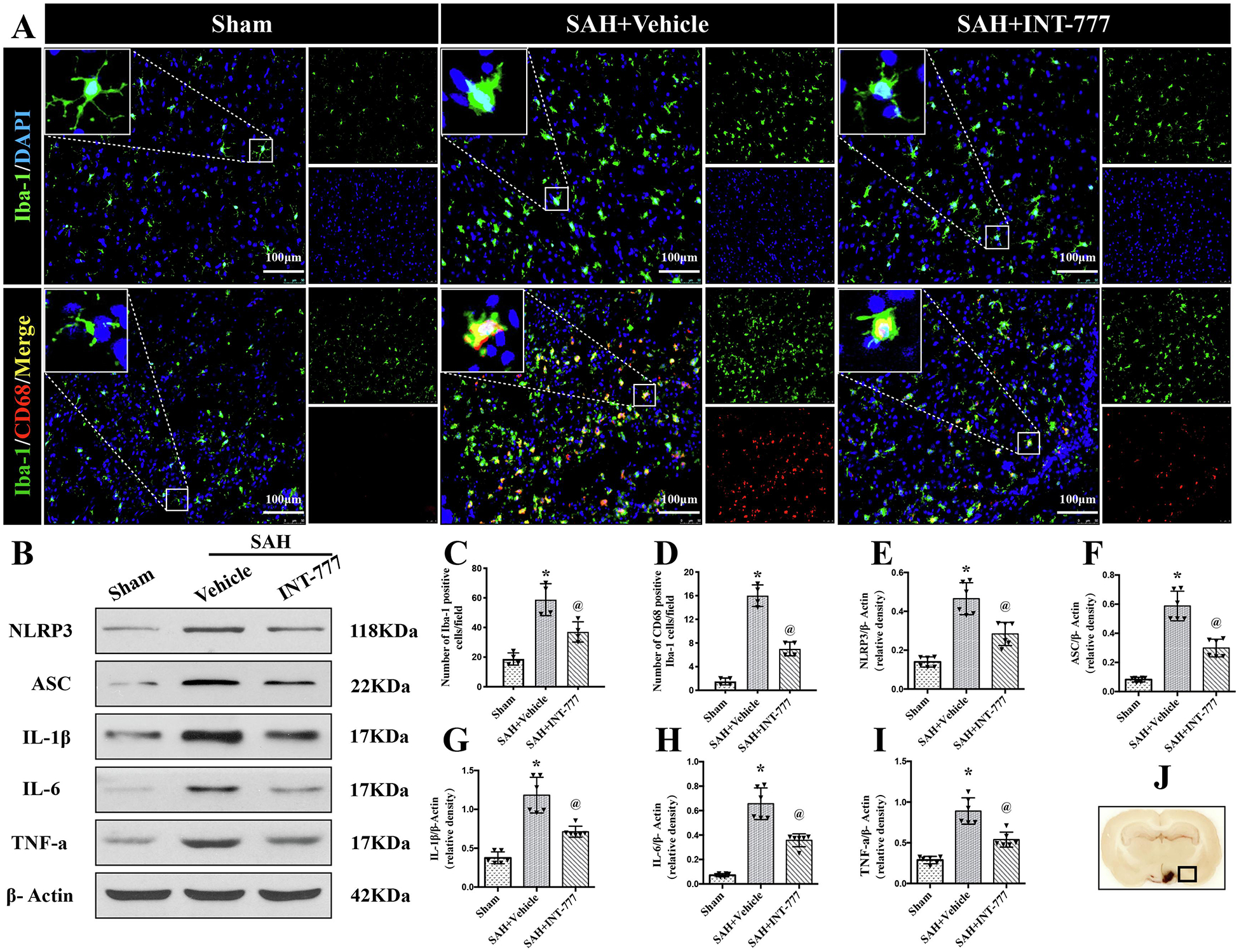
Figure 3. Intranasal administration of INT-777 reduced microglial NLRP3-ASC inflammasome activation-mediated neuroinflammation within ipsilateral hemisphere at 24 h after SAH.

(A) Representative immunofluorescence microphotographs of microglia (Iba-1, green) and CD68 (red)-positive activated microglia in the ipsilateral basal cortex for sham, SAH+Vehicle, and SAH+INT-777 groups. Nuclei were stained with DAPI (blue). Scale bar=100 μm (J) A small black squares in the coronal section of brain indicated the area used for microphotographs. (C-D) Quantitative analysis of Iba-1-positive and CD68-positive microglia. n=4 per group. (B, E-I) Representative western blot bands and densitometric quantification of NLRP3, ASC, IL-1β, IL-6, and TNF-a in the ipsilateral hemisphere at 24 h after SAH, n=6 per group. Vehicle: 10% dimethyl sulfide. Data was represented as mean ± SD. *P<0.05 vs. Sham group; @P<0.05 vs. SAH+Vehicle group.
