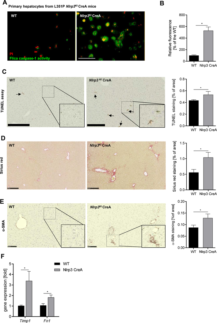
Figure 4. Hepatocyte-specific Nlrp3 mutant mice (L351P Nlrp3KICreA) showed increased hepatocyte caspase-1 activation and increased fibrogenesis.
(A) Flica-caspase-1 activity (green) and PI (red) stained Nlrp3KICreA and WT primary hepatocytes (scale bar: 100 μm). (B) Quantification of caspase-1 positive cells (%) normalized on WT hepatocytes. (*p< 0.05 vs. WT hepatocytes). Immunohistological stainings and quantification of livers from WT and Nlrp3KICreA mice for (C) TUNEL positive cells (scale bar: 250 μm), (D) Sirius red collagen disposition (scale bar: 250 μm), and (E) α-SMA protein (scale bar: 100 μm). (F) mRNA expression of Timp1 and Fibronectin 1 (Fn1) in Nlrp3KICreA livers. Two groups were analyzed by Studentś T-test (*p< 0.05).

