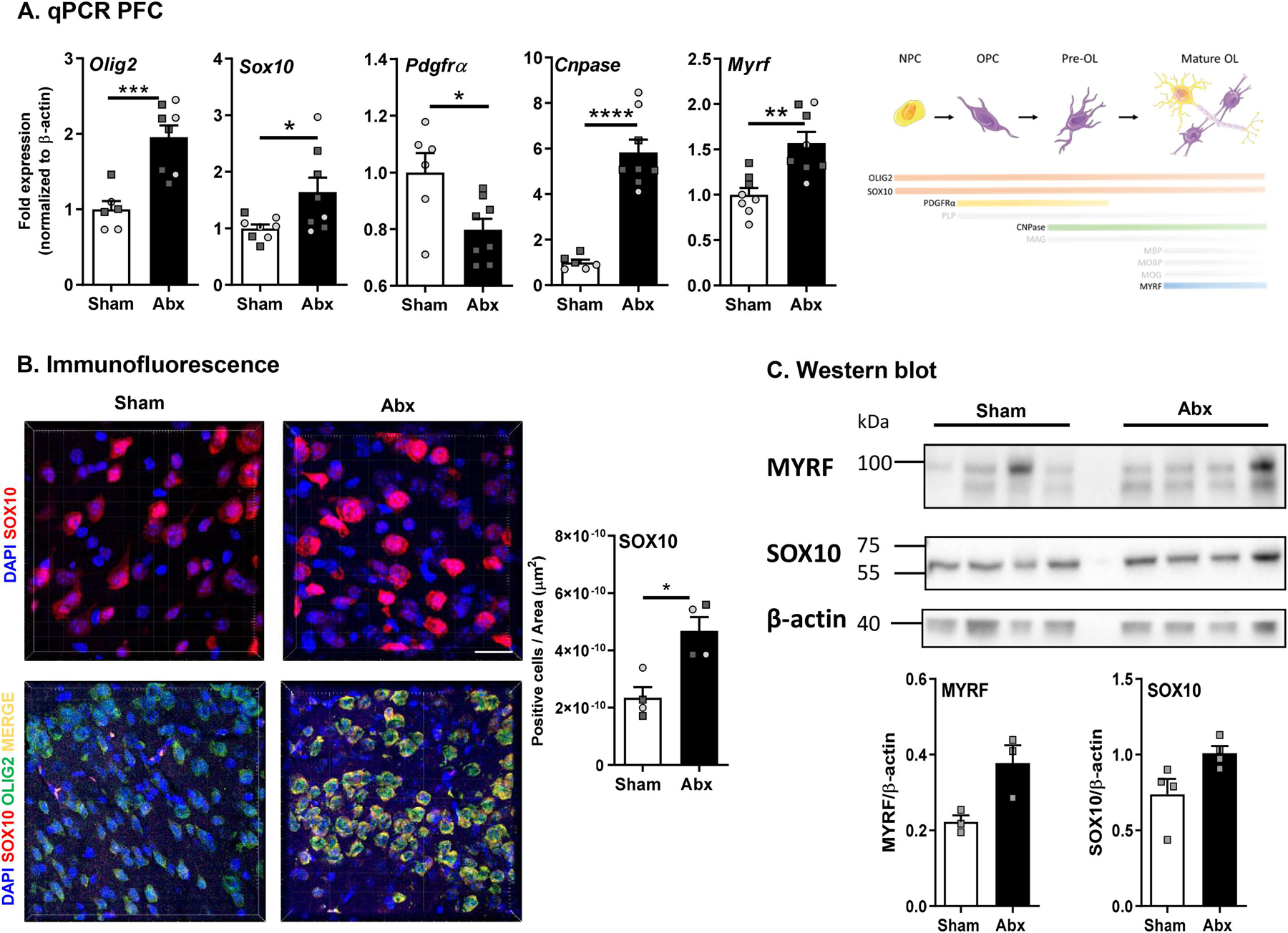
Fig. 5.

Neonatal Abx administration leads to maturation and differentiation of oligodendrocytes. (A) mRNA expression of oligodendrocyte lineage-specific markers in the pre-frontal cortex (PFC) (N = 7–8). (B) Immunofluorescence for SOX10 (63x) expression in the PFC (N = 4). DAPI in blue and SOX10 in red. Scale bar 20 µm. Unpaired T-test *p < 0.05; co-localisation staining of OLIG2 and SOX10 (40x) in the PFC region (N = 4). DAPI in blue, SOX10 in red, OLIG2 in green, merge in yellow). Scale bar 50 µm. (C) Western blot and densitometric analysis for MYRF, SOX10 and β-actin (N = 5). Females denoted as light grey circles, and males as dark grey squares. Student’s T-test *p ≤ 0.05.
