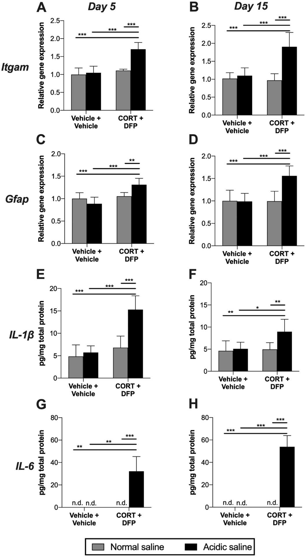
Figure 4. Levels of neuroinflammatory markers in DRG.

Corticosterone (CORT) or vehicle was administered via the drinking water for 7 days to mimic high physiological stress. On the final day of CORT treatment, a single dose of diisopropyl fluorophosphate (DFP) or vehicle was administered. Seven days later, rats received a single injection of normal (pH = 7.2) or acidic saline (pH = 4.0) into the left gastrocnemius muscle. Left L4/5 DRG were collected 5 or 15 days later. Expression levels of Itgam (CD11b) were determined at (A) day 5, (B) and day 15, as well as Gfap at (C) day 5, (D) and day 15. Protein levels of IL-1β were assayed at (E) day 5 (F) and day 15. IL-6 levels were also assayed at (G) day 5 (H) and day 15, using the lower limit of detection for statistical analysis of undetectable values. *P < 0.05, **P < 0.01, ***P < 0.001. N = 6–8/group. Data are mean ± SD.
