
Figure 3:
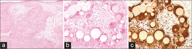
The histological examination revealed (a) a nerve sheath tumor constituted by well-differentiated Schwann cells, characterized by nuclear palisades consistent with Verocay bodies (H and E, ×40). The microscopic study at ×100 highlighted foci of foreign body reaction (H and E in b) with uniformly sized, grey-yellowish microspheres phagocytized by multinucleated giant cells (immunostained with CD68 in c).