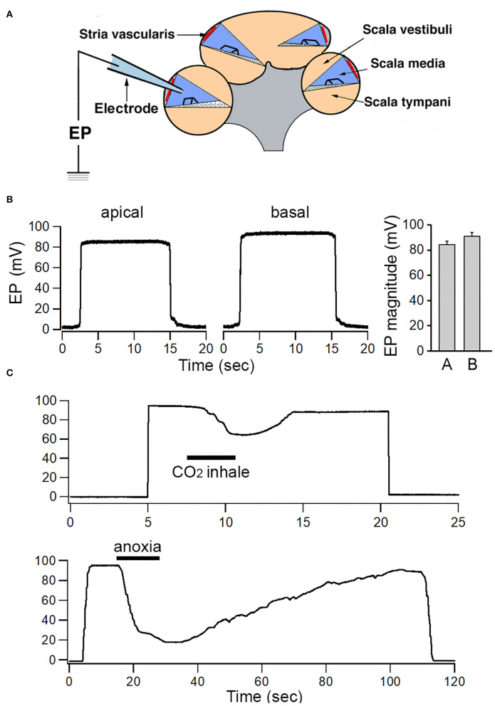
Figure 1.
Recording of EP from apical and basal turns of the cochlea. (A) Schematic view of electrode path for recording EP through lateral wall approach. (B) Examples of EP recorded from apical and basal turns. Mean and SD of EP recorded from eight apical and eight basal turn locations are shown in the right panel. (C) Change in EP magnitude in response to temporal CO2 inhalation and anoxia.

