Figure 3.
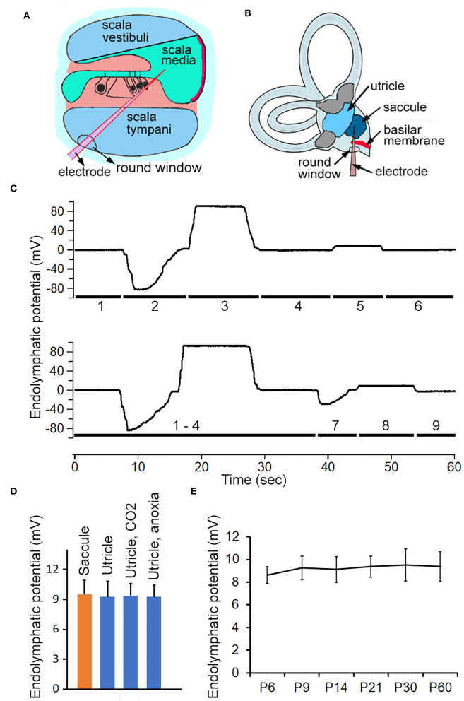
Recording ELP from cochlea, saccule and utricle in adult and neonatal mice. (A) Schematic view of electrode path for recording EP through round window approach. (B) Electrode path for recording ELP from saccule through round window. (C) Representative examples of changes in recorded potential as micropipette advancing from scala tympani, through basilar membrane, and into scala media and scala vestibuli, and finally into saccule or utricle. The number underneath the response indicates the location where the response was recorded. 1: scala tympani. 2: region of organ of Corti. 3: scala media. 4: scala vestibuli. 5: saccule. 6: scala vestibuli. 7: utricular macula. 8: utricle. 9: withdrawing the electrode (into scala vestibuli would return the potential to zero or a slightly negative potential. (D) Mean magnitude and SD of ELP recorded from saccule and utricle (n = 8 for either). Utricular ELP magnitude after inhalation of CO2 and anoxia is also presented. No statistical significance was found among them. (E) Development of ELP of saccule. Mean and SD are presented (n = 8).

