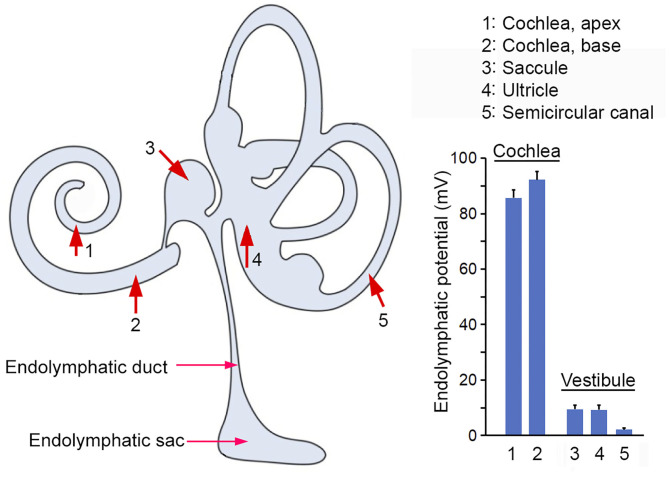
Figure 6.
ELP recorded from the inner ear. Red arrows and numerical numbers mark the location where ELP was measured. The magnitude of ELP at different endolymphatic compartments in the inner ear is shown in the right panel. Schematic drawing in the left panel was modified from Kim et al. (2011) with permission.

