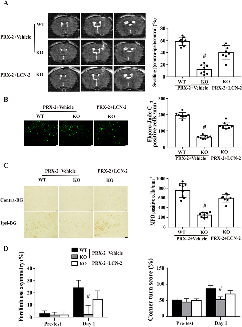
Figure 5: Exogenous LCN-2 enhances PRX-2 induced brain damage in male LCN-2 KO mice.
Male LCN-2 KO mice received an intracerebral co-injection of PRX-2 with recombinant mouse LCN-2 protein or vehicle. Another control group of male WT mice received PRX-2 with vehicle. A) Representative T2 MRI at day 1 showing brain swelling by PRX-2. Brain swelling was quantified by ventricular compression. B) Fluoro-Jade C staining of degenerative neurons in the ipsilateral basal ganglia (BG) at day 1. C) Neutrophil infiltration in the ipsilateral BG at day 1. D) Forelimb use asymmetry and corner turn test. Values are mean ± SD, n=8, #P < 0.01 vs. the PRX-2+Vehicle-WT and PRX-2-KO group by one-way ANOVA with Tukey’s post-hoc test.

