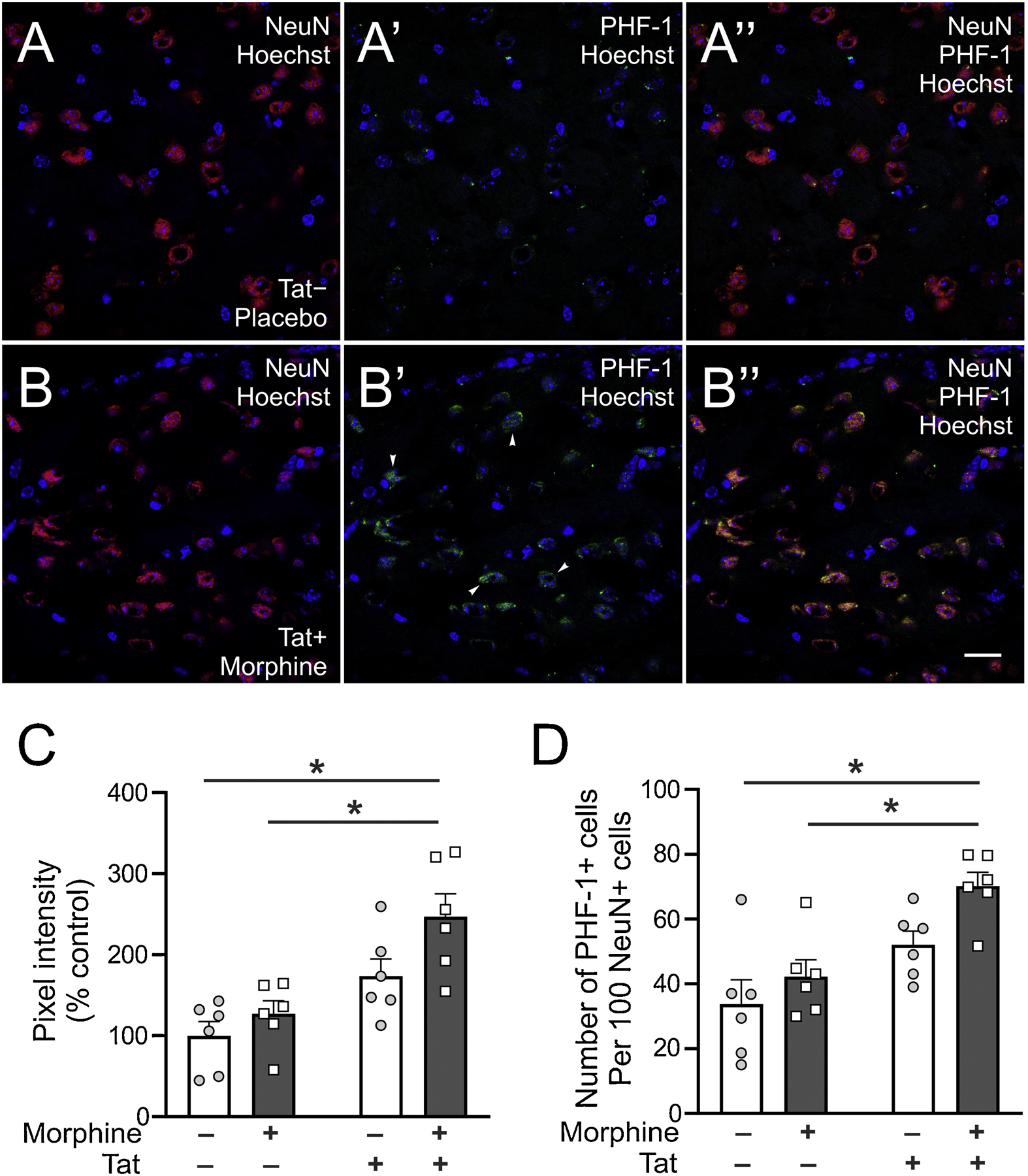
Fig. 2.

Striatal PHF-1 immunoreactivity. (A and B) Representative photomicrographs (63×) showing increased PHF-1 immunoreactivity in the striatum of Tat− control mice (A) and morphine-treated Tat+ (B) mice. Scale bar; lower panel = 20 μm. Arrowheads show PHF-1 stained cells. (C) Striatal PHF-1 immunoreactivity was increased in mice co-exposed to Tat and morphine compared to Tat− mice irrespective of treatment. (D) Neuron-specific expression of PHF-1 was increased in mice co-exposed to Tat and morphine compared to Tat− mice irrespective of treatment. Data are expressed as the mean ± SEM. * indicates planned contrasts that showed differences using Tukey’s post hoc test (p < 0.05). NeuN: neuronal marker, Hoechst: nuclear counterstain, PHF-1: Paired helical filament-1 antibody for detection of tau neurofibrillary tangles phosphorylated at serine 396/404. n = 6/group.
