Figure 1.

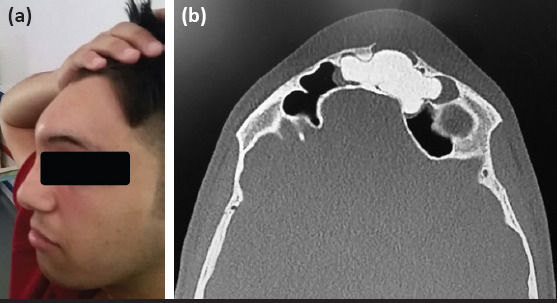
(a) The appearence of swelling, approximately 2x2 cm, in the frontal area. (b) The mass eroding the left frontal sinus anterior wall on paranasal sinus tomography

(a) The appearence of swelling, approximately 2x2 cm, in the frontal area. (b) The mass eroding the left frontal sinus anterior wall on paranasal sinus tomography