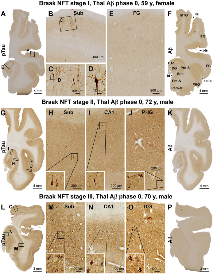
Figure 1.
Verification and Braak staging of primary age-related tauopathy (PART) in postmortem human brains. Shown are micrographs of adjacent temporal lobe sections immunolabeled for phosphorylated tau (pTau) and β-amyloid (Aβ) from representative cases examined in the current study, with the patient’s biometrics (age and sex) and image panel arrangements indicated. The left and the middle panels show the regional distribution and morphology of pTau-immunoreactive neuronal profiles in the three cases with Braak stages I (A–E), II (G–J), and III (L–O) neurofibrillary tangle (NFT) lesions, respectively. The right panels (F,K,P) show the lack of Aβ deposition in the sections immunolabeled with the monoclonal antibody 6E10 in all the cases (scored as Aβ Thal phase 0). In the first case (A–E), a small population of pyramidal neurons located around the border of CA1 and subiculum exhibited strong pTau immunoreactivity in the somata and dendritic arbors, with light labeling seen in their axons (A–C), while no labeling is seen in the temporal neocortex (TC; E). In the second case (G–J), pTau-labeled neuronal somata and processes occur in the CA1, transentorhinal, and entorhinal areas (G–I) and occasionally in the basal TC (J). In the third case (L–O), pTau-labeled neuronal somata and processes are densely present in the limbic areas (L–N) and also frequently seen over the temporal neocortical gyri (O). col-S, collateral sulcus; DG, dentate gyrus; FG, fusiform gyrus; HF, hippocampal fissure; ITG, inferior temporal gyrus; MTG, middle temporal gyrus; its, inferior temporal sulcus; ots, occipito-temporal sulcus; PHG, parahippocampal gyrus; Sub, subiculum; Para-S, parasubiculum; Pre-S, presubiculum; Pro-S, prosubiculum. Scale bars and enlargements of local areas are as marked in the panels.

