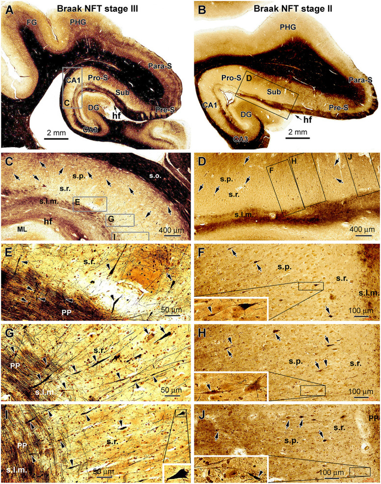
Figure 2.

Representative silver-stained micrographs illustrating dystrophic dendritic profiles in the hippocampal formation in the brains with PART. Shown are low- and high-magnification views of the labeling in the temporal lobe sections from two cases (cases #22 and #16 in Table 1) with tauopathy at Braak stages III (A,C,E,G and I) and II (B,D,F,H and J), respectively. The framed areas in the panels are enlarged as indicated. The arrows point to examples of silver-stained somata of CA1 and subicular pyramidal neurons, while the arrowheads point to examples of silver-stained dendritic swellings. The dystrophic dendritic parts are featured by segmented fusiform expansion along the course of the labeled dendritic processes, which are mostly located over the stratum radiatum (s.r.). Some dendritic swellings occur in the stratum lacunosum-moleculare (s.l.m.), intersecting with the fine axons of the perforant pathway (C–J). In some cases, one silver-stained apical dendrite has repeated swellings along its proximal to distal continuation (G,I). The swelling can be also seen at the proximal segments of dendrites (F,H). Other abbreviations are as defined in Figure 1, and scale bars are as indicated.
