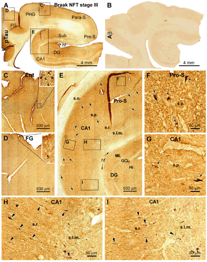
Figure 4.

Cross-validation of the existence of dendritic dystrophy in the hippocampal formation with phosphorylated tau (pTau) immunohistochemistry. (A) Case #22 in Table 1: low-magnification view of pTau labeling over the hippocampal formation, the entorhinal cortex, and part of the TC. The boxed areas (C–E) are enlarged to show the distribution of pTau-immunolabeled neurons in the hippocampal formation (E) and entorhinal cortex (C) and occasionally in the neocortical fusiform gyrus (D). (B) Lack of cerebral β-amyloid (Aβ) deposition in this case. The framed areas in (E) are further enlarged (F–I) to illustrate the somata (as pointed by arrows) of immunoreactive hippocampal and subicular pyramidal neurons. (H,I) Segmental fusiform swellings (as pointed by arrowheads) of the dendritic processes in the s.r. and stratum lacunosum-moleculare (s.l.m.). Additional abbreviations are as defined in Figures 1, 2. Scale bars in individual panels are as indicated.
