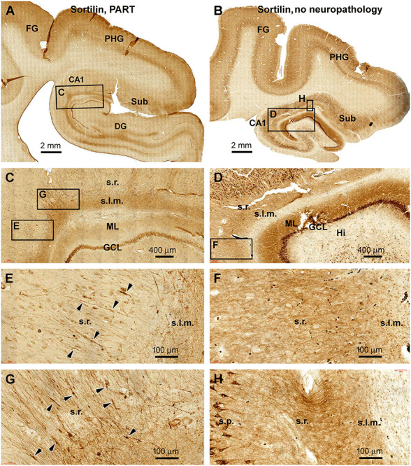
Figure 5.

Observation of dendritic dystrophy in hippocampal formation in primary age-related tauopathy (PART) case in sortilin immunohistochemistry. (A) Sortilin labeling in the section adjacent to the one shown as Figure 4A (case #22 in Table 1). (B) Labeling in the comparable region in a case without cerebral pTau or Aβ pathologies (case #32). The framed areas are enlarged as indicated. In the PART case (C,E,G), fusiform swellings (as pointed by arrowheads) are seen on the immunolabeled dendritic processes in the s.r. In contrast, no swelling is found on the dendritic processes in the control case (D,F,H). Other abbreviations are as defined in Figure 1. Scale bars are as indicated.
