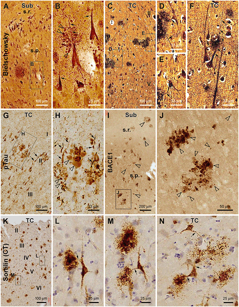
Figure 6.

Characterization of plaque-associated axonal and dendritic pathologies. (A–F, cases #27 and 28 in Table 1) Silver-stained neuritic plaques in the subiculum and TC as indicated. (G–J, case #27) Phosphorylated tau- and β-secretase 1-immunoreactive neuritic profiles. (K–N, case #25) Neuronal profiles and extracellular plaques in the TC immunolabeled with the sortilin C-terminal antibody. Axonal dystrophic neurites (DN; as pointed by open arrowheads) appear as swollen globular profiles which occur in clusters and as small isolated sphericles as well. Dendritic neurites (as pointed by arrows) are elongated processes with local swelling or thinning, some of which are connected to the labeled neuronal somata. Panel arrangement, lamination, and scale bars are as indicated. I–IV, cortical layers. Additional abbreviations are as defined in Figures 1, 2.
