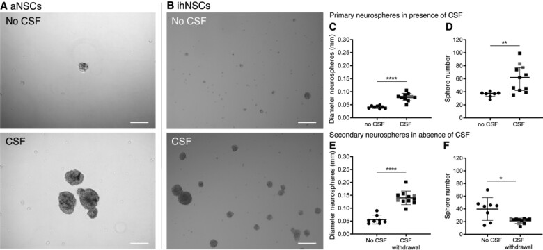
Figure 3.
Increased neural stem cell (NSC) proliferation upon treatment with ventricular CSF. (A) Representative image of primary adult NSC (aNSC) culture of donor NBB 2012-066. aNSCs treated with 25% pooled post-mortem ventricular CSF were observed to form more and larger neurospheres than untreated aNSCs after 5 days in culture. (B) Representative image of immortalized human NSC (ihNSC) culture without and with CSF of donor NBB 2011-096. ihNSCs treated with 25% post-mortem ventricular CSF (n = 11 wells treated with CSF of individual donors) form larger and more neurospheres compared to untreated cells (n = 8 untreated wells) after 5 days. (C, D) The ihNSC neurosphere diameter (two-tailed t-test with Welch’s correction, t13.39 = 8.13, P < 0.0001) and sphere number (Mann–Whitney U test, U = 6, P = 0.0018) were quantified after 5 days of CSF treatment. (E, F) Seven days after CSF withdrawal, passaged ihNSC neurosphere diameter (two-tailed t-test, t17 = 7.877, P < 0.0001) and number (two-tailed t-test with Welch’s correction, t7.558 = 2.887, P = 0.0216) were quantified. In grey, the data points belonging to the wells treated with CSF of Alzheimer patients are indicated; exclusion of these data points did not change the statistical results. Data expressed as mean ± standard deviation in C, E and F, and as median ± interquartile range in D; *P < 0.05, **P < 0.01, ****P < 0.0001; Scale bars = 100 μm.

