Figure 5.
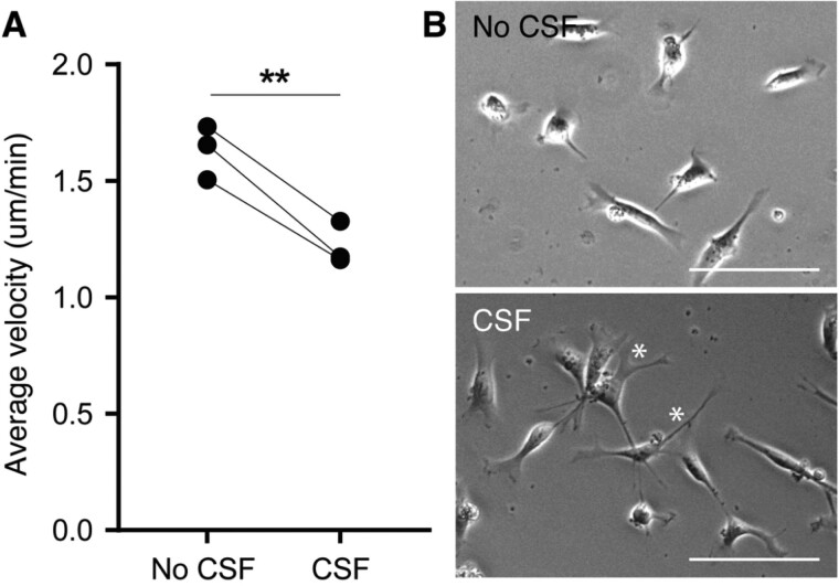
Decreased cell motility and changed morphology of ihNSCs after ventricular CSF pre-treatment. (A) Average velocity of cells pre-treated with CSF (n = 3 culture replicates) was significantly lower compared to untreated cells (n = 3 culture replicates) (paired t-test, t2 = 10.17, P < 0.0095). Per-culture replicate, five cells were traced per five locations per condition. Data are expressed as mean ± standard deviation. (B) Representative phase-contrast pictures of untreated ihNSCs and ihNSCs pre-treated with CSF; the former showing rounder cells, the latter showing a more elongated morphology with more protrusions (*). Scale bars = 100 μm.

