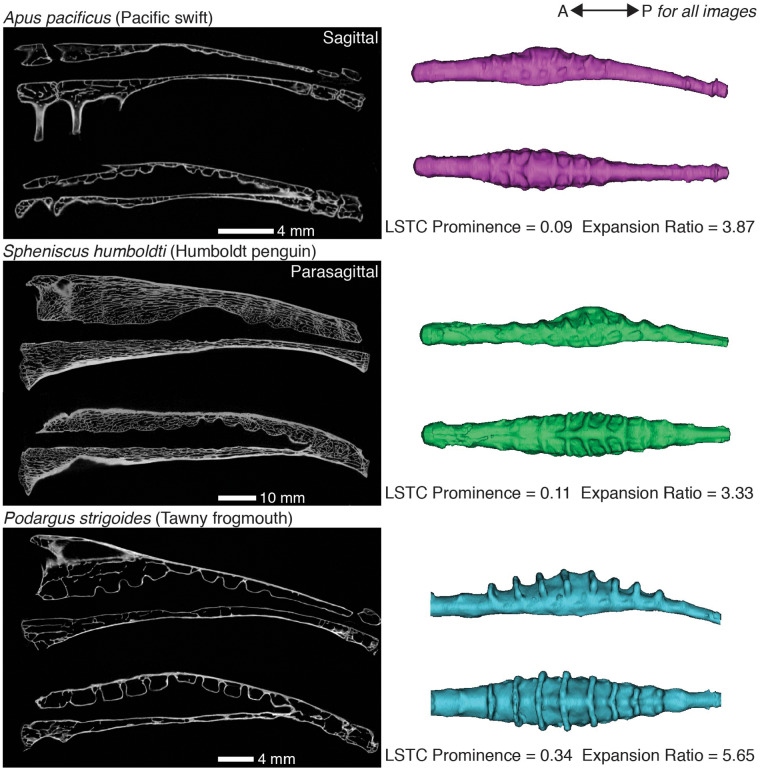
Fig. 4.
The morphology of the synsacral vertebral canal varies across bird species. These are sagittal and parasagittal sections from μ-CT scans of the synsacra of three bird species paired with lateral and dorsal views of the corresponding vertebral canal endocasts. Although A. pacificus and S. humboldti have similar values for the metrics we calculated, the shape of their endocasts differ, particularly in the orientation of the LSTCs (see dorsal views of endocasts). We calculated much higher values of both metrics for P. strigoides than for the other two species shown here, and the endocast of P. strigoides exhibits correspondingly visually prominent LSTCs and central expansion.

